Knee meniscal tear

What is a meniscus?
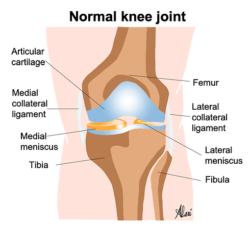
The meniscus is a C-shaped structure made of a special type of cartilage (called hyaline cartilage). There are two menisci in each knee (a lateral meniscus at the outer aspect of the knee joint and a medial meniscus at the inner aspect of the knee joint). The menisci are very important for normal knee function and stability. They deepen the surfaces between the bones at the knee joint and connect to various other knee structures, thus providing stability to the joint. They also act as shock absorbents and cushions to the joint, reducing the pressure on the articular cartilage. The articular cartilage is the important layer of tissue that lines the surface of the bones at the joints and gets worn off in conditions like knee arthritis.
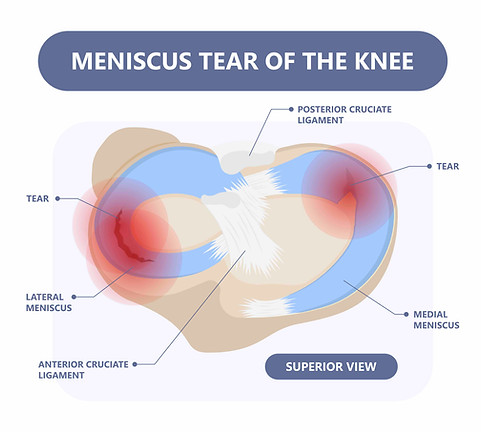
The menisci are essential in reducing the stress upon the joint and protecting the knee joint from injury. The meniscus is susceptible to injury, particularly if exposed to a sharp force secondary to sudden twisting or rotation movement. This can happen in sports activities, and this type of injury is usually referred to as (an acute meniscal tear). Tears can also happen without a specific injury, especially in the older population, and this type is referred to as (chronic degenerative meniscal tear).
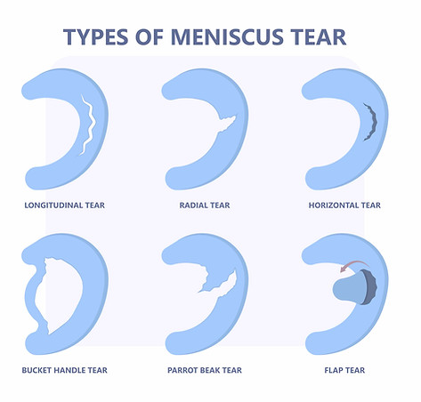
The medial meniscus bears the most significant pressure during weight bearing and is, therefore, most susceptible to injury. There are different types and classifications of meniscal tears, including horizontal, longitudinal and radial tears (according to their direction within the meniscus substance) and displaced and non-displaced meniscal tears (according to the presence or absence of meniscal tissue). The type of the tear will significantly affect how it is treated.
What are the features of a meniscal tear?
The signs and symptoms usually associated with a meniscal tear include:
-
Sudden, sharp pain after an unexpected twisting or rotation. This can happen during sports or other activities.
-
The pain is felt more at the inner aspect of the knee in the presence of a medial meniscal tear and more on the outside in the presence of a lateral meniscal tear. However, if there are tears in both menisci, the pain can be widespread all over the knee.
-
Giving way (feeling that the knee might suddenly collapse under you).
-
Knee locking. This typically happens when there is a displaced meniscal tear.
-
Localised or generalised knee joint swelling. Localised knee swelling can be due to a small pocket of fluid next to the tear (a para meniscal cyst). Generalised swelling can be due to knee joint inflammation and fluid build-up. This can happen because the torn meniscus is not properly cushioning, leading to increased stress upon the joint.
What other conditions that can mimic a meniscal tear?
A meniscal tear can be confused
-
Injury to the ligaments around the knee joint (called the collateral ligaments)
-
Pes anserine bursitis (can be confused with a medical meniscal tear).
To read more about other causes of pain/swelling at the inner or outer aspect of the knee which can be confused with a medial or lateral meniscal tear respectively, please see our articles.

Horizontal tear to the posterior horn of the meniscus
What is a parameniscal cyst?
A para meniscal cyst is a focal build-up of fluid adjacent to the meniscal tear. This can happen if the meniscal tear act as (a one-way valve) allowing fluid to get from the knee into this pocket but not allowing the fluid to go back. It is usually felt as a small area of swelling at the outside or back of the knee. Clinical diagnosis of this can be challenging. Imaging (ultrasound and MRI) is very useful in assessing this and confirming the diagnosis.
How to diagnose a meniscal tear?
When a meniscal tear is suspected clinically, imaging is usually needed to establish the diagnosis. MRI is the most sensitive modality for assessment. It will assess the menisci and other important structures like the articular cartilage for any injury. Visualisation of a meniscal tear on ultrasound can be challenging. A para meniscal cyst can be usually seen on ultrasound. More importantly, ultrasound would be very useful to guide any injection treatment, if indicated.
Is a knee injection useful for a meniscal tear?
An injection can be useful for managing a meniscal tear, but the decision is usually taken after careful clinical assessment and obtaining an opinion from an orthopaedic surgeon. Injection treatment could include steroid/corticosteroid, hyaluronic acid and platelet-rich plasma (PRP) injections. Injection treatment in patients with meniscal tears is usually indicated in certain circumstances:
-
Patients with meniscal tears who are trying to avoid surgery.
-
Degenerative type meniscal tears. These tend to happen gradually with no definite history of injury. Sometimes, these are associated with degenerative knee joint changes, which usually also benefit from injection therapy.
-
Failure of conservative management like rest, anti-inflammatory tablets, physiotherapy and muscle strengthening exercise.
Most importantly, ultrasound-guided knee joint injections should be done under ultrasound guidance. There is significant evidence that when injections are done under ultrasound guidance, they result in better outcomes in pain relief, duration of effect and function improvement. This is because ultrasound guidance allows for accurate placement of the needle and, thus, delivery of the medicine to the exact site of pain. Ultrasound can be used to inject the desired medicine into the knee joint. It can also guide the aspiration procedure if there is a sizeable para meniscal cyst that can be painful and tense. Ultrasound-guided aspiration of a para meniscal cyst means inserting a small needle into the cyst using ultrasound guidance to extract the fluid from the cyst. Please see our article to read more about para meniscal cysts.

Ultrasound-guided corticosteroid injection
Corticosteroid (cortisone) is a well-established anti-inflammatory medicine for knee pain and inflammation. Cortisone is usually mixed with a short-acting local anaesthetic (numbing medicine) to provide quick pain relief. To find out more, please read our article about knee joint steroid injections
Ultrasound-guided hyaluronic acid injection
Hyaluronic acid can be useful in the management, especially if knee osteoarthritis is associated. It has a dual anti-inflammatory and shock-absorbent effect. It aims to simulate the joint's natural lubrication, allowing movement to occur in a low-friction environment. Hyaluronic acid injections are not known to have adverse effects on the articular cartilage (unlike cortisone) and can be repeated if symptoms return. We usually use high molecular weight Hyaluronic acid preparations (like Ostenil Plus and Durolane). These are proven to be the most effective and need a single injection only. To find out more, please read our article about Hyaluronic acid injections
Ultrasound-guided platelet-rich plasma (PRP) injections
Ultrasound-guided PRP injections are usually used if there is associated knee joint osteoarthritis. To find out more, please read our article about Ultrasound-guided platelet-rich plasma (PRP) injections
Summary
The menisci are important structures for knee joint stability and function. A meniscal tear can result in significant pain, swelling and instability. Imaging plays important role in establishing the diagnosis and guiding further management. Ultrasound can be very useful if any injection treatment is indicated.
Recent posts


Related posts


Categories
Knee conditions and treatments

Specialist Consultant Musculoskeletal Radiologist Doctor with extensive experience in image-guided intervention
To book a consultation
Call us on 020 8050 9885 or Book online
The Musculoskeletal Ultrasound & Injections clinic
Brentford, TW8 9DR
